Continuous Bubbling in Water Seal Chamber
- Introduction
- Aim
- Definition of terms
- Indications for insertion
- Insertion of a chest drain
- Chest drain assessment & management
- Specimen collection
- Chest drain dressings
- Removal of chest drain
- Complications and Troubleshooting
- Family Centred Care
- Companion Documents
- Evidence Table
Introduction
Chest drains also known as under water sealed drains (UWSD) are a drainage system of three chambers consisting of a water seal, suction control and drainage collection chamber. UWSD are designed to allow air or fluid to be removed from the pleural cavity, while also preventing backflow of air or fluid into the pleural space. This allows for the expansion of the lungs and restoration of negative pressure in the thoracic cavity. Appropriate chest drain management is required to maintain respiratory function and haemodynamic stability. Chest drains may be placed routinely in theatre, PICU and NICU; or in the emergency department and ward areas in emergency situations. Some patients will have Redivac drains inserted, these are different from a UWSD. Please refer to Pleural and Mediastinal Drain Management after Cardiothoracic Surgery guideline.
Aim
To provide a clear guide for nursing staff to promote the safe, correct and competent management of the UWSD in the ward setting. This clinical guideline is written from the perspective of a cardiac and renal ward management; however, it also outlines management from the perspective of other speciality areas where care may differ.
Definition of terms
- Chylothorax: Collection of lymph fluid in the pleural space
- Haemothorax: Collection of blood in the pleural space
- Pleural effusion: Exudate or transudate in the pleural space
- Pneumothorax: Collection of air in the pleural space
- Tension Pneumothorax: Air builds up in the pleural space and forces a mediastinal shift leading to decreased venous return to the heart and lung collapse/compression causing acute life-threatening respiratory and cardiovascular compromise.
Indications for Insertion of a Chest Drain
- Chylothorax
- Pleural effusions
- Pneumothorax
- Post cardiac surgery
- Haemothorax
Insertion of a Chest Drain
Please see the Chest Drain Insertion CPG below:
- Chest Drain (Intercostal Catheter) Insertion Clinical Practice Guideline.
RCH access only: See Aseptic Technique Policy and Procedure
Chest Drain Assessment & Management
Start of shift checks
- Ensure that there is emergency equipment at bedside including:
- At least two drain clamps per drain (For use in emergency only)
- Two suction outlets - x1 chest drain and x1 for airway management
- Auscultate the chest
- Assess the chest tube and system tubing (i.e. for kinks, dislodgement etc) as well as the drain dressing to ensure it is intact and for any signs of infection
- Check that the drain is anchored appropriately
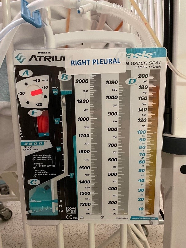
- Labelling of each drain tube in accordance with EMR documentation (See image below)
- Drain is on suction (i.e Red bellow out) and that the amount of suction correlates with the medical team order on EMR (See image below for labelling of each chamber)
- Assess for any leakages or movement in water chamber
- Check that the drain in not clamped (unless ordered by medical staff).
- Significant risk of tension pneumothorax development if clamped

- Significant risk of tension pneumothorax development if clamped
UWSD Labelling
It is imperative that the UWSD is labelled in a way that is clear and visible. This is extremely important when removing the drain to ensure the correct drain is removed. Please refer to the image below for the correct labelling of an UWSD. The criteria for correct labelling includes the following:
- How - Clear, visible writing (Dark font and capital letters) on a label that can be complete stuck down
- What – Include the position (e.g. Pleural, mediastinal etc) and side (e.g. right, left)
- Where – Top of the USWD & avoid covering any measurements / numbers
- Who & when – Labelling should be completed by the Cardiac theatre nurses / surgeons post operatively prior to transfer of the patient back to the ward. Should a UWSD require changing post operatively on the ward, the responsibility of labelling lies with the direct care giver (e.g. Nurse changing the UWSD or bedside nurse).

Patient Assessment
Vital signs
- Auscultate chest at shift commencement
- Routine four hourly observations including assessment of respiratory effort
- If the patient is on an opioid infusion continuous monitoring is required and hourly cardiorespiratory observations (HR, SpO2, BP, RR) should be documented
For ward areas
- On insertion of chest drain monitor and document on EMR flowsheets patient observations (HR, SpO2, BP, RR, respiratory effort and temperature):
- 15 minutely for 1 hour
- 1 hourly for 4 hours then 1-4 hourly as indicated by patient condition
Drain insertion site and dressing assessment:
- Check that the dressing is clean and intact
- Assess for any ooze, bleeding or signs of infection/inflammation every hour and document this within EMR:
- Small amounts of ooze can be a normal presentation post operatively, however if any abnormal presentations are observed: Notify the medical team, consider taking and sending swabs of the site and take a clinical photo and upload onto EMR
- Observe that the sutures remain intact and secure (particularly long term drains where sutures may erode over time)
Skin integrity:
- Assess skin at a minimum four hourly for pressure injuries particularly where the tubing is in contact with the skin. Effective anchoring below the insertion sites will assist with preventing this.
UWSD Unit and Tubing
- Ensure all connections between chest tubes and drainage unit are tight and secure
- Assessment of chest tube and system tubing should occur at the beginning of the shift and every hour throughout the shift. Tubing should have no kinks or obstructions that may inhibit drainage
- Never lift drain above chest level. The unit and all tubing should be below patient's chest level to facilitate drainage. Ensure the unit is securely positioned on its stand or hanging on the bed
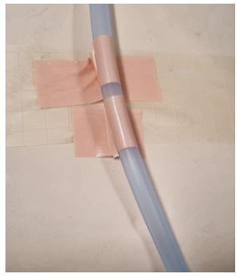
- Drain tubing should always be secured to patient bed to prevent accidental removal and anchored to the patient's skin to prevent pulling of the drain (See image below)
- Ensure the water seal is maintained at 2 cm at all times
Pain
- Ensure that patients with an UWSD have appropriate and regular pain relief (IV opioid infusion, PCA etc). A CPMS referral should also be in place and also consider appropriate administration of pain relief prior to mobilisation, physiotherapy sessions or other activities that involve movement etc
- Pain assessment should be conducted and documented four hourly unless more frequently warranted
Suction
- Ensure that the suction is on and ensure that the "red bellow" is all the way out
- If suction is required orders should be written by medical staff and documented within the "Orders" tab on EMR. Suction on the drainage unit should be set to the prescribed level, please see below. Suction is not always required as it may lead to tissue trauma and prolongation of an air leak in some patients.
- -5 cmH20 is commonly used for neonates
- -10 cmH20 to -20 cmH20 is usually used for children
Drainage
- Every hour, the UWSD tubing should be "tilted and tipped" in order to ensure the fluid within the tubing accumulates within the chamber
- Milking of chest drains using roller clamps is only to be done with written orders from medical staff. Milking drains creates a high negative pressure that can cause pain, tissue trauma and bleeding
Volume
Document hourly the amount of fluid in the drainage chamber (for each drain if multiple present) in the Fluid Balance flowsheet on EMR
Assess the cumulative total of drainage output and notify medical staff of the following:
- A sudden increase in amount of drainage
- Greater than 5mls/kg in 1 hour
- Greater than 3mls/kg consistently for 3 hours
- Significantly reduced output over an extended length of time or if a drain with ongoing loss suddenly stops draining
- Blocked drains are a major concern for cardiac surgical patients due to the risk of cardiac tamponade
If the chamber tips over and blood has spilt into next chamber, simply tip the chamber up to allow blood to flow to original chamber
Colour and Consistency
Colour and consistency of drainage should be documented hourly along with documentation of volume amounts. If there is a change (eg. Haemoserous, bright red, serous, chylous/creamy) notify medical staff immediately
- Consider taking sample of drainage to assess for chylothorax (refer to Specimen Collection). See Chylothorax Management Guideline
Air Leak (bubbling)
- The water seal chamber should be assessed every hour for any potential air leaks. An air leak will be characterised by intermittent bubbling in the water seal chamber when the patient with a pneumothorax exhales or coughs.
- The severity of the leak will be indicated by numerical grading on the UWSD (1-small leak 5-large leak)
- Continuous bubbling of this chamber indicates large air leak between the drain and the patient. Check drain for disconnection, dislodgement and loose connection, and assess patient condition. Notify medical staff immediately if problem cannot be remedied.
- If suspected or confirmed leakage, clamp drain tubing closest to patient and escalate accordingly and document on Fluid Balance Flowsheet on EMR
Oscillation (Swing)
- The water in the water seal chamber will rise and fall (swing) with respirations. This will diminish as the pneumothorax resolves. Assess and document on Fluid Balance Flowsheet on EMR
Other Considerations
- Referral to a physiotherapist should be made to enhance chest movement and prevent a chest infection and mobilisation and transferring should be encourage as appropriate where possible
- Parent education on managing potential accidental dislodgement should be discussed
Patient Positioning
- Patients who are ambulant post operatively will usually have fewer complications and shorter lengths of stay. If a patient is on strict bed rest or is an infant, regular changes in position should be encouraged to promote drainage, unless the patient's clinical condition prevents doing so
Patient Transport
- If the patient needs to be transferred to another department or is ambulant, the suction should be disconnected and left open to air
- Remember that during transport, the tubing should not be clamped and that the UWSD should remain below chest level
- Clamps must not be used on the patient for transport because of the risk of tension pneumothorax
- When transporting the patient, be weary of tubing getting caught in the bed and utilise a "cart/trolley" where possible for ease of transport especially for multiple drains. Remember to remove anchor from bed before mobilising the patient.
Specimen Collection
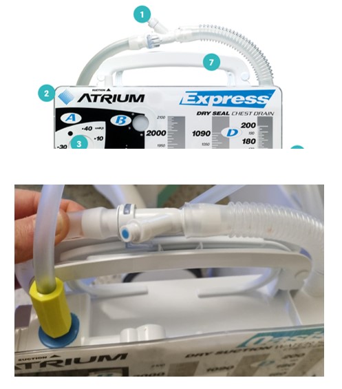
Collect drainage specimens for culture through the needless sampling port located by the in-line connector.
Equipment Required
- Specimen container
- Alcohol swab
- 10ml syringe
- Dressing pack
- Gloves
- Eye Protection
Procedure:
1. Wait for the fluid to collect in a loop of the tubing
2. Perform hand hygiene, then don gloves & eye protection
3. Clean the sampling port ( See feature labelled as 1 in the image below) with an alcohol wipe and leave to dry for 20 seconds
4. Temporarily clamp the tubing above where the fluid has collected
5.Connect a 10ml Luer lock syringe to the sampling port and aspirate the fluid out of the tubing. Do not pierce tubing/sampling port
6. Place fluid in sterile specimen container
7. Once the syringe is disconnected remove all clamps and kinks
8. Perform hand hygiene
Chest Drain Dressings
Dressing Change
- Dressings should be changed:
- Every seven days unless otherwise indicated
- If they are no longer dry and intact, or signs of infection
- Infected drain sites require daily changing, or when wet or soiled
- Changes are a risk for accidental drain removal. Avoid unnecessary changes
- Dressings should ensure drain is secure
- Dressings should allow site visibility and prevent pressure on skin
Equipment Required
- Dressing pack
- Chlorhexidine solution (Yellow or blue)
- Gloves
- Two small occlusive tegaderm dressings
- Split gauze
Procedure:
1. Perform hand hygiene and then don gloves
2. Remove old dressing
- Preferably get someone who is not sterile to don latex gloves and remove the dressing
3. Clean the drain site using the appropriate chlorhexidine solution:
- Use standard aseptic technique with body temperature sterile normal saline
- If the wound is showing signs of being infected, contaminated or soiled use the yellow solution (Aqueous Chlorhexidine 0.15% w/v cetrimide 15%)
4. Once the site is dry apply two layers of split gauze in the direction where the split facing up and it wraps around the drain. Another two layers of split gauze should then be placed on top of the first layer of gauze in the opposite direction (split facing down).
5. Cover gauze using hypafix TM (approx two pieces, enough to cover the gauze completely)
6. Remove gloves and perform hand hygiene
For cardiac surgical patients with drains inserted intraoperatively: Ensure dressing does not communicate with sternotomy dressing or wound
Anchoring drain tubing
- Apply ComfeelTM or similar to protect fragile skin from 'tag' of tape
- Use "sleek" TM or "hypafix" TM to secure the drain tube (see image below). When securing and folding the sleek around the tubing, ensure that the tubing isn't secured flat down and that there is a "gap of tape" between the comfeel TM and tubing
Removal of Dressings
- To remove dressing when placed flat against the skin - Lift corner from the skin and slowly stretch the in dressing in a motion that is parallel to the skin
- To remove semi permeable dressing placed in a sandwich position
- Hold the corners of the dressing on either side of the drain and pull them away from each other; this should create a pocket around the drain
- Peel each of the dressings away from each other until you reach patient skin
- Slowly stretch the rest of the pressing in a motion that is parallel to the patient's skin to remove the rest of the dressing.
- To remove semi permeable dressing placed in a sandwich position
Changing the Chamber
-
Indications
- The chest drain chamber needs to be replaced when it is ¾ full or when the UWSD system sterility has been compromised e.g. accidental disconnection.
-
Equipment Required
- New UWSD
- Dressing pack
- Gloves
- Eye Protection
Procedure
1. Perform hand hygiene
2. Use personal protective equipment to protect from possible body fluid exposure
3. Using an aseptic technique, remove the unit from packaging and place adjacent to old chamber
4. Prepare the new UWSD as per manufacturer's directions supplied with drain
5. Ensure patients drain is clamped to prevent air being sucked back into chest
6. Disconnect old chamber by holding down the clip on the in-line connector to pull the tubing away from the chamber.
7. Insert the tubing into the new chamber until you hear it click.
8. Unclamp the chest drain
9. Check drain is back on suction
10. Place old chamber into yellow infectious waste bag and tie
11. Perform hand hygiene
Removal of Chest Drains
Important considerations for drain removal
- There must be written documentation for drain removal by medical staff in EMR specifying which drain/s are to be removed
- The responsibility of the proceduralist removing drains is to check the patient's post-operative x-ray prior to drain removal
- Chest X ray Must be checked by the proceduralist before any chest drain removal and after removal. The X ray is then reviewed by surgical team to follow up if further management is required.
- Drain labelling must be physically checked by proceduralist prior to removal to ensure removal of correct drain/s. Consult cardiac surgeon for further clarification.
Indications
- Drainage diminishes to little or nothing
- Absence of an air leak (pneumothorax)
- No evidence of respiratory compromise
- Chest x-ray showing lung re-expansion
Equipment required
- Dressing trolley with Yellow Infectious waste bag attached
- Dressing pack (sterile towel, sterile gauze)
- Sterile Gloves
- Appropriate skin cleaning solution for procedure
- Neonates <1500 grams - Chlorhexidine irrigation solution 0.1% (Blue Solution)
- For all other patients - Aqueous Chlorhexidine 0.15% w/v cetrimide 15% (Yellow Solution)
- Steristrips
- Suture Cutter
- Band Aids
- Clamps
- Eye Protection
- Occlusive dressing
- Sharps container
Patient and pre-procedure preparation
- Ensure patient is fasted, has been administered adequate and age-appropriate pain control, sedation (fast appropriately) and distraction therapy (see Procedural Pain Management Guideline)
- Consider environment e.g. treatment room or privacy screens if in ward area and the involvement of Child Life Therapy if required
- Ensure you have enough staff to assist with the procedure & ensure that the patient is monitored throughout the procedure
- Heparin infusions for cardiac patients should not be discontinued prior to drain removal
- Always remove pacing wires (cardiac patients) before removing the drains and ensure that the drains are unclamped during the removal of wires
Procedure
1. Perform hand hygiene
2. Opening dressing pack and add sterile equipment and 0.9% saline
3. Don disposable gloves
4. Remove all dressings around the area
5.Clamp drain tubing and ensure suction is disconnected – double check with assisting nurse. If there are multiple drains in-situ, clamp all drains before removal. Once the required drains are removed, unclamp remaining drains
6. Remove disposable gloves, perform hand hygiene and don sterile gloves
7. Place sterile towel under tubes
8. Clean around catheter insertion site and 1-2cm of the tubing with age appropriate skin cleaning solution
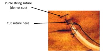
9. If purse string present (cardiac patients) unwind in preparation for assistant to tie
10. Remove suture securing drain (ensuring purse string suture not cut)

11. Instruct patient to exhale and hold if they are old enough to cooperate; if not, time removal with exhalation
12. Pinching the edges of the skin together, remove the drain using smooth, but fast, continuous traction
13. The assistant pulls purse string suture closed as soon as the drain is removed, tying 2 knots and ensuring the suture is not pulled too tight. Cut tails of suture about 2cm from knot
14. If there is no purse string present remove drain and quickly seal hole with occlusive dressing (i.e. TegadermTM)
15. Apply steristrips over insertion site if needed
16. Remove and discard equipment into a yellow infectious waste bag and tie
17. Perform hand hygiene
Post Procedure Care
- Attend to patients comfort and sedation score as per procedural sedation guideline
- CXR should be performed post drain removal, ideally 2 hours post (max 4 hours)
- Patients in PICU may wait until routine daily CXR if clinically well
- Clinical status is the best indicator of re-accumulation of air or fluid. CXR should be performed if patient condition deteriorates
- Monitor vital signs closely (HR, SpO2, RR, Respiratory effort and BP) on removal and then every hour for 4 hours post removal, and then as per clinical condition
- Document the removal of drain in the LDA flowsheet in EMR
- Remove sutures four days post drain removal
- Dressing to remain in situ for 24 hours post removal unless contaminated
Complications and Troubleshooting
- Pneumothorax - Signs and symptoms include:
- Decreased SpO2, increased respiratory effort , diminished breath sounds, decreased chest movement, complaints of chest pain, tachycardia or bradycardia, hypotension
- Notify medical staff & request an urgent CXR
- Prepare for insertion/repositioning of chest drain
- Ensure drain system is intact with no leaks, or blockages such as kinks or clamps
Bleeding at the drain site
- Don gloves
- Apply pressure to insertion site
- Place occlusive dressing over site
- Notify medical staff
- Check Coagulation results
- Check drain chamber to ensure no excessive blood loss
Infection of insertion site
- Notify medical staff
- Swab wound site
- Consider blood cultures
Accidental disconnection of system
- Clamp the drain tubing at the patient end. Clean ends of drain and reconnect. Ensure all connections are cable tied. If a new drainage system is needed cover the exposed patient end of the drain with sterile dressing while new drain is setup. Ensure clamp is undone when problem has resolved.
- Check vital signs and consider getting a CXR
- Alert medical staff
Accidental drain removal
- Apply pressure to the exit site and seal with steristrips. Place an occlusive dressing over the top
- Check vital signs, consider the need for a CXR and alert medical staff
- The incident must be documented in the VHIMS by the nurse attending to procedure
Purse string cut or not present
- Small bore drains such as pigtails do not require purse strings. Simply apply an occlusive dressing.
- For large bore drains:
- Pinch or apply pressure to the exit site
- Apply steristrips to close exit site and cover with an occlusive dressing
- Notify the responsible medical team to review patient and consider need for a suture
- The incident must be documented in the VHIMS by the nurse delegated to remove the drain.
Unable to remove chest drain
- If the drain is unable to be removed with reasonable traction being applied, notify the responsible surgical team
- If unsafe to be removed on the ward, the patient might have to go to theatre and have it removed under general anaesthetic
Retained drain during removal
- If the tube fractures during drain removal and remnants of the tubing are left within the patient contact the treating team immediately (Initiate MET call as needed or if patient becomes unstable).
- An urgent chest x-ray should be conducted.
- The patient should be prepared for theatre e.g. keep fasted
- The whole drain unit should be kept in the patient's room until surgical review and will need to be kept for collection to enable quality review.
- The piece of drain tubing that remains in the patient will also be kept once surgically removed to allow for appropriate follow up of the incident's cause.
- The incident must be documented in the VHIMS by the nurse delegated to remove the drain.
- Please refer to Surgical Drains (Non Cardiac) guideline & RCH policy for Missing or Non-Intact Drains
Family Centred Care
- Explain purpose of chest drain to family and when it is likely to be removed
- Discuss the need for pain relief for the child to be comfortable enough to move and participate in physiotherapy
- Encourage parental involvement with mobilising, sitting out of bed and assisting with chest physiotherapy to aid faster recovery.
- Multidisciplinary care e.g. Physiotherapy referral
Companion Documents
- Chest Drain Insertion Guideline
- Cardiac Surgical Wound Dressing
- Atrium Drains Website
- Procedural pain management
- Chylothorax Management CPG
- PICU Outreach Service
- Surgical Drains (Non Cardiac) guideline
Evidence Table
- Evidence table for Chest Drain Management
Please remember to read the disclaimer
The development of this nursing guideline was coordinated by Charmaine Russell, CNS, Koala, and Emily Mohd Faizal, RN, Koala, and approved by the Nursing Clinical Effectiveness Committee. Updated October 2022.
Source: https://www.rch.org.au/rchcpg/hospital_clinical_guideline_index/Chest_drain_management/


0 Response to "Continuous Bubbling in Water Seal Chamber"
Post a Comment